[tab name=”The Case”]
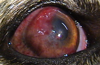
This little Jack Russell terrier is polydipsic and polyuric and losing weight into the bargain. You look into his eyes for no particular reason (apart from your fascination with ophthalmology of course!) and this is what you see. What is the connection with his systemic signs?
[/tab]
[tab name=”David’s view”]
These apparent bubbles are vacuoles that are the earliest sign of diabetic cataract. All (or at least most!) of the books say that the classic diabetic cataract is one that suddenly presents as a mature white cataract like this one below.
 But that’s, to my mind at least, just because diabetic dogs don’t get shown to an ophthalmologist until they are suddenly blind. In fact the vast majority of diabetics have vacuoles where water influx into individual lens fibres has occured way before they suddenly progress to being fully opaque. Now the question is what can be done to stop them progressing. If we could inhibit the aldose reductase enzyme converting the glucose to sorbitol which sucks in the water, no diabetic dog need be blind. A topical preparation shown to have this effect is not available currently though has been reported (Kador PF, Webb TR, Bras D, Ketring K, Wyman M. Topical Kinostat ameliorates the clinical development and progression of dogs with diabetes mellitus. Vet Ophthalmol. 2010;13:363-8) and we have good evidence for similar results with an oral preparation, Ocuglo, but more of that when our study is complete!
[/tab]
[end_tabset]


You must be logged in to post a comment.