A differential list of one?


[tab name=”The Case”]

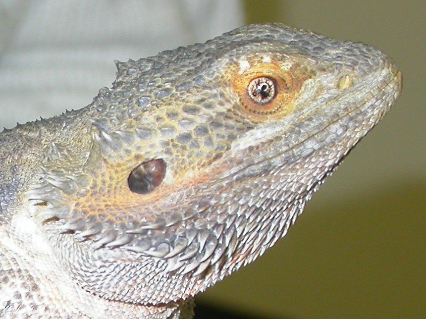
A 9 year old labrador with droopy lower and upper eyelids, a somewhat enophthalmic globe with third eyelid protrusion and a miotic pupil normally means just one thing to me. But this dog had a somewhat painful eye as well which made me think of a second explanation of these signs. What was I thinking of? 

[/tab]
[tab name=”David’s view”]

 This triad of signs is classic of Horner’s syndrome where the ‘fight flight’ nerve dilating the pupil, opening the upper lid and bringing the eye forward, is not functioning as it should. But the inclusion of some ocular discomfort made me realise, for the first time in nearly 25 years on the job, that a corneal ulceration with intraocular inflammation could just about present with the same signs of miosis and globe retraction with blepharospasm.

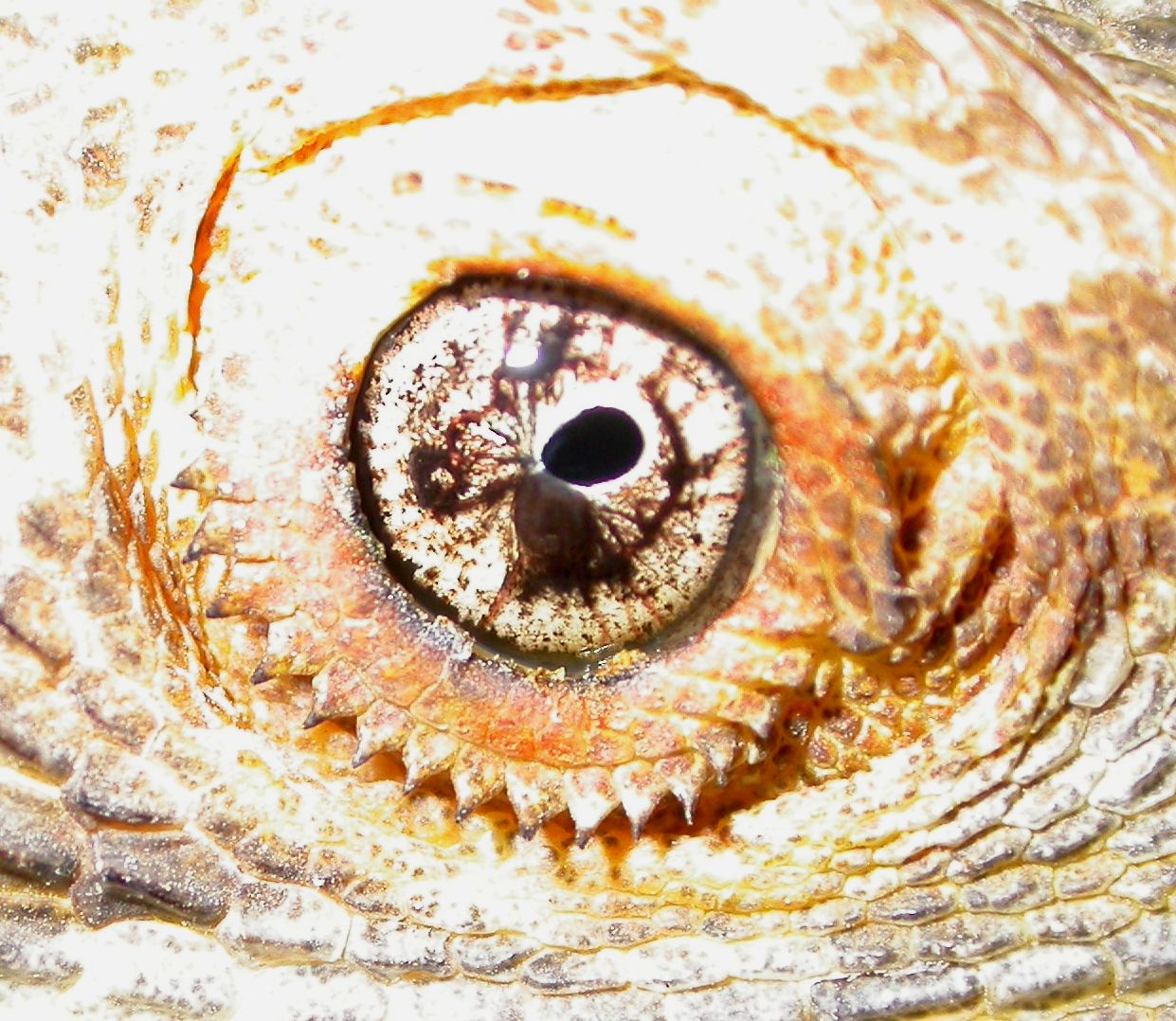
 The way to be sure of the diagnosis was using 0.1% phenylephrine and the two pictures below show the dog ten and twenty minutes after topical installation of this sympathomimetic agent.


The signs start to resolve within ten minutes and are fully (one might say over-fully!) ameliorated in twenty. This suggests a third order neuron defect as there is denervation hypersensitivity at the motor end plate. A closer look at the eye showed no signs of corneal ulceration or inflammation although interestingly tonometry showed a somewhat lower intraocular pressure which could indicate a mild uveitis.

The definitive diagnosis of what was causing the defect came with MRI showing increased signal intensity characteristic of an inflammatory lesion in the middle ear on the ipsilateral side (the big white blob if you are an imaging ignoramus as I am!). Hopefully resolution of this otitis media, maybe with a bulla osteotomy, will solve the problem but I’ll leave that up to the surgoens! 

Many thanks to Abi at Dick White Referrals for this great scan!

[/tab]
[end_tabset]

Posted in Cases | Leave a comment

Will this cure cataracts?


[tab name=”The Case”]


 This is n-acetyl carnosine, an antioxidant developed by Dr Mark Babizhayev who claims that his product has beneficial effects when applied to eyes with cataract. I was interested in this product around ten years ago and conducted some preliminary research into its use in 30 dogs, which we published in the peer reviewed journal Veterinary Ophthalmology. The big question is does this compound work to resolve cataract, rendering this sort of eye visual again?

[/tab]
[tab name=”David’s view”]

 The simple answer from our research (available at http://davidlwilliams.org.uk/resources/file0256.pdf) is that in mature blinding cataracts the eyedrop appears to have no beneficial effect whatsoever. In the very earliest stages of lens opacification there may be some slowing of cataract development but if a dog has a severe cataract the only treatment, and a very effective one at that, is phacoemulsification surgery, as shown below. If you are reading this as a dog owner and your pet is becoming blind, or is already blind, visit a veterinary ophthalmologist to discuss surgery before inflammatory changes or retinal detachment render surgery less likely to be successful.


[/tab]
[end_tabset]

Posted in Cases | 2 Comments

What do we do with this?


[tab name=”The Case”]

 My good friend Dominic Alexander e-mailed me this picture, not because he wanted a diagnosis, but rather to know what I would suggest as a treatment plan.


 What would your diagnosis and treatment be?

[/tab]
[tab name=”David’s view”]

 This is an iris cyst. In the horse, unlike the dog, they are generally attached to the iris and, if they grow in size, they can cause the horse to spook as they move around.  Here is another view of this one.

 In the USA these are generally lasered (see for example Gemensky-Metzler AJ, Wilkie DA, Cook CS. The use of semiconductor diode laser for deflation and coagulation of anterior uveal cysts in dogs, cats and horses: a report of 20 cases. Vet Ophthalmol. 2004:360-8), but here a simple needle inserted in through the ventral cornea under sedation and a local nerve block can serve to deflate the cyst quite well.

[/tab]
[end_tabset]

Posted in Cases | Leave a comment

Blindsight?

 [tab name=”The Case”]

 This five year old labrador has dilated pupils without much of a pupillary light reflex and a retina that looks just like the picture in the textbook for generalised progressive retinal atrophy with an overly reflective tapetum and narrow vessels.

But the dog can see quite happily to traverse a maze of chairs in the waiting room and follows a dropped ball of cotton wool with no problem whatsoever. Is your presumptive diagnosis wrong?

[/tab]
[tab name=”David’s view”]

 Here’s another picture of the dog’s retina with the flash attenuated somewhat so the vessels are easier to see.

 No – your diagnosis is correct. The dog has early gPRA, but the key early feature of this is nyctalopia, night blindness. Turn off the room lights and the dog is blind as a bat – or actually more blind, as it doesn’t have echo-location! It will be interesting to see how rapidly the phototopic or day vision reduces. The condition the dog has is known as rod-cone degeneration, as the rods die first followed by the cones, with their demise probably accelerated by the higher oxygen levels given that there are no rods left to use the oxygen. We are hoping that using a mixture of dietary anti-oxidants we can slow down this cone death and thus maintain day vision for a considerable time. If you are interested in reading more about this my article on oxidative stress and the eye  in Veterinary Clinics of North America, though rather old by now, may be of interest – find it at: http://davidlwilliams.org.uk/resources/file0261.pdf.

[/tab]
[end_tabset]

Posted in Cases | Leave a comment

Is this a melanoma?


[tab name=”The Case”]

 This cat has been referred with a possible iris melanoma.

 What is your opinion?

[/tab]
[tab name=”David’s view”]

 It’s black and it looks a bit sinister for sure, but this is ectropion uveae, migration of the pigmented epithelium of the posterior face of the iris through to the anterior face.  A much less severe example is seen in the fellow eye, with a focal anterior cataract in for good measure!

[/tab]
[end_tabset]

Posted in Cases | Leave a comment

The ultimate botox!


[tab name=”The Case”]

 This poor labrador was afflicted with botulism and spent a week on a ventilator before recovering well as seen here.

While paralysed a carbomer gel was assiduously applied to both eyes, but treatment ceased when she was able to breathe again. A few days later I was called to see her and closer inspection showed these changes to her ocular surface.

What is happening and what should we do?

[/tab]
[tab name=”David’s view”]

 There is a grey hazy corneal oedema in a linear streak in both eyes and a break-up of the reflection from the camera flash, showing a defect in the tear film or the corneal epithelium or both. There was indeed ulceration and an evaporative dry eye even though her Schirmer tear test was over 20mm/min in each eye. These changes were associated with a failure to blink adequately. We used a cross-lnked hyaluronan gel (Sentrx Remend) with excellent results as can be seen below. There is still some oedema but the epithelium is perfectly healed, as seen by the crisp clear flash reflection.

The case shows the importance of protecting the ocular surface whenever blinking is defective, for instance during long surgical operations or during coma in the ICU.

[/tab]
[end_tabset]

Posted in Cases | Leave a comment

A big bad black hole!


[tab name=”The Case”]

 I was asked to look at this black labrador as he was extremely hypertensive with a suspected phaeochromocytoma. I thought I might see the same sort of retinal changes as we detect in a cat, though to my mind they occur much less frequently in hypertensive dogs. But what should I see but this?! What do you reckon it is and what would you do next?


[/tab]
[tab name=”David’s view”]

 I don’t think there is much of a differential list here – this is a spot diagnosis and a big black spot one at that! Choroidal melanomas are rare in dogs but look just like this and I can’t think of anything else that does though this is my first one in 25 years of ophthalmology! An ultrasound scan shows that the tumour doesn’t invade much further than the choroid. While the few papers in the literature on the tumour in dogs do document metastasis, this is generally late on in tumours that were much more advanced when dioagnosed than this one.

Sorry that this isn’t the best ultrasound scan in the world but shows the ‘collar stud’ appearance classic of these neoplasms.  Note that there is a flat retinal detachment visible on the scan and numerous small bullae of detachment classic of hypertensive retinopathy around the tumour and further afield on the retinal photo. They occur in the otherwise normal fellow eye below, baving said that this is much less severe than a feline hypertensive retinopathy at a similar blood pressure of nearly 300mmHg. If you can explain why cats and dogs are so different in this regard do please tell!!

[/tab]
[end_tabset]

Posted in Cases | 3 Comments

Bad eye in a beardy!

 [tab name=”The Case”]

This bearded dragon has an unusual right eye compared with his left but doesn’t seem to be affected by it. What might be happening here?

[/tab]
[tab name=”David’s view”]

Here’s where a bit of magnification comes in useful!

The pupil is constricted and at its lower boundary lies a pigmented mass. This could be an iris cyst or a relatively benign melanoma but I find it difficult to be sure. Ultrasonography tended to suggest a cystic structure but the minute size of the mass (less than a millimetre in diameter)made interpretation difficult.  Given the small size of the eye and the lack of effect on the animal at present, surgery to remove the mass  would probably cause far more ocular pathology than it removed, so for the moment we are going to watch and wait. If it enlarges over the next month or two laser photocoagulation would be my best therapeutic bet!

 [/tab]
[end_tabset]

Posted in Cases | Leave a comment

An alien in the eye?!


[tab name=”The Case”]

This appaloosa horse was referred to me with a blind but pain-free eye which seems to be enlarged.

It was impossible to see through the pupil so we use ultrasound to investigate the intraocular pathology, and what should we find but an alien in the posterior segment!

What on earth are we seeing?!

 [/tab]
[tab name=”David’s view”]

 The eye has, as is common in this breed, experienced intraocular inflammation with the pupil becoming completely closed.

The intraocular pressure then rose increasing the size of the eye. An ultrasound scan at a slightly different angle shows an obliterated anterior chamber with the iris and cornea touching, a secondarily luxated lens and a retinal detachment. The ‘alien’ is actually a bunched up fold of retina viewed at an unusual angle.

The horse is not at all concerned about the eye, even though the intraocular pressure is 56mmHg, so we are not planning on doing anything apart from watching the other eye (currently completely normal) very carefully. And there I was hoping to publish a report of the first UFO identified in an animal eye!

[/tab]
[end_tabset]

Posted in Cases | Leave a comment

The chelonian eye in health and disease

The Chelonian Eye in Health and Disease – a review of tortoise and turtle ocular anatomy and physiology together with disease aetiology, pathogenesis and treatment.

Link here or in the preview panel below:

Testudo-paper.pdf


Posted in Publications | Leave a comment