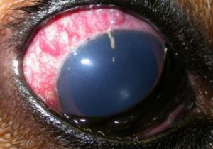
A Clinical Case from the Archives : 31/07/2005

[tab name=”The Case”]This ocular appearance is seen on routine examation of a 14 year old mixed breed dog which the owners report as having no obvious visual deficit. What are you seeing here?[/tab][tab name=”David’s view”]The dense white opacities in the anterior vitreous here are asteroid hylaosis. These bodies are hydroxylapatite consisting of calcium and phosphate according to Komatsu and colleagues (Fine structure and morphogenesis of asteroid hyalosis.
Med Electron Microsc. 2003 36:112-9) whileWinler and Lunsdorf (Ultrastructure and composition of asteroid bodies. Invest Ophthalmol Vis Sci. 2001 42:902-7)also note the importance of proteoglycans and hyaluronate in the vitreous in the biomineralisation process occurring in these older eyes. Moss and colleagues showed asteroid hylosis to be present in 1.2% of the aged human population in Beaver Dam, Wisconsin as shown here(Asteroid hyalosis in a population: the Beaver Dam eye study. Am J Ophthalmol. 2001 132:70-5) but we have, as yet, no idea of the prevalence in the older pet dog population.[/tab][end_tabset] 

Posted in Cases | Tagged , | Leave a comment

A Clinical Case from the Archives : 30/07/2005

[tab name=”The Case”]My seven year old son reckoned this dog had been watching the telly too long – Jack knew that ‘watching the box can give you square eyes’! What are this bulldog’s problems?[/tab][tab name=”David’s view”]Well Peggy Sue’s other eye is a triangle not a square but look at the conjunctival hyperaemia and corneal neovascularisation as well as the lids – the dog has KCS with a Schirmer tear test of under 5 in each eye as well as some lid entropion. She needs topical tear replacement, cyclosporine and lower lid surgery too.[/tab][end_tabset] 

Posted in Cases | Tagged , , | Leave a comment

A Clinical Case from the Archives : 30/07/2005

[tab name=”The Case”]This dog has a localised area of redness in the left eye. Are there other signs that aid you in your diagnosis? Might there be a systemic disaese associated with this condition?[/tab][tab name=”David’s view”]Note the scleral swelling here too. This is scleritis. In man the systemic association is with a more generalised connective tissue disease be it rheumatoid arthritis or Wegener’s granulomatosis though we don’t seem to see this connection in the dog. This animal also has an epitheliotropic lymphoma – lymphoma masquerading as apparent scleritis is certainly seen in man (Dorey Orbital lymphoma misdiagnosed as scleritis. Ophthalmology. 2002 109:2347-50 for example), though this is the first case I’ve seen in the dog.[/tab][end_tabset] 

Posted in Cases | Tagged , | Leave a comment

A Clinical Case from the Archives : 27/07/2005

[tab name=”The Case”]This Basset hound comes to you with a two day history of a red painful eye and the owner’s suspicion that vision on that side may not be all it should be. What’s the diagnosis, how would you confirm that and what emergency treatment would you give to improve the condition?[/tab][tab name=”David’s view”]This is glaucoma, caused because of dysplasia of the pectinate ligament of the iridocorneal angle in both eyes confirmed by tonometry showing an elevated intraocular pressure (iop) and gonioscopy of the angle in the still healthy fellow eye showing an abnormal pectinate ligament. Professor Gelatt has recently shown that over 5% of Bassets in the USA have glaucoma (Prevalence of the breed-related glaucomas in pure-bred dogs in North America.
Vet Ophthalmol. 2004 7:97-11) making it one of the worst affected breeds there and I guess here in the UK too. In the old days we would have given mannitol as an iv drip, but these days topical prostaglandin, in the form of Latanaprost or a similar agent, constricts the pupil, as you can see here and lowers the pressure in minutes. Here the iop was 37mmHg, lowered to 24mmHg in twelve minutes and under 20 over the next hour, by the drug opening up the unconventinal drainage route through the choroid which bypasses the iridocorneal angle altogether. Magic! Doctor Wilis and colleagues have produced a useful review of new topical anti-glaucoma agents in their 2002 paper ‘Advances in topical glaucoma therapy’ appearing in Veterinary Ophthalmology 5:9-17.[/tab][end_tabset] 

Posted in Cases | Tagged , | Leave a comment

A Clinical Case from the Archives : 26/07/2005

[tab name=”The Case”]A good friend of mine from college days has just sent me some pictures of 2-3 day old calves – both Friesian and Angus cross animals – developing these white ocular lesions. They seem to resolve in a few days but no necessarily completely. What is happening and why?[/tab][tab name=”David’s view”]This is corneal oedema – sometimes there is so much excess water in the corneal stroma that the cornea swells and becomes deformed with a keratoconus. This can occur with malignant catharral fever or with phenothiazine administration but the cause here is still a mystery – plesae give me an e-mail if you have an idea![/tab][end_tabset] 

Posted in Cases | Tagged , | Leave a comment

A Clinical Case from the Archives : 22/07/2005

[tab name=”The Case”]This 8 week old puppy is presented to you with a sore ey. What is likely to be going on here?[/tab][tab name=”David’s view”]Eversion of the lid edges as shown here, demonstrates that the swelling is related to the meibomian glands and is most likely to be an ocular manifestation of juvenile cellulitis. Hot water compresses and topical anti-inflammatory medication will be helful, but complete resolution is likely to be best acheieved with a short course of systemic steroid.[/tab][end_tabset] 

Posted in Cases | Tagged , | Leave a comment

A Clinical Case from the Archives : 22/07/2005

[tab name=”The Case”]This terrier was given a lamb bone to chew at the weekend. Early the next week the owner reports he had a ‘puffy looking eye’ but that this calmed down with systemic antibiotic. A week later the eye looked like this. What might be going on and what diagnostic steps would you take?[/tab][tab name=”David’s view”]This eye, as they say, is ‘hot’! The intraocular pressure was 45mmHg and an ultrasound shows the classic ‘sea gull’ sign of retinal detachment with fluid or blood behind the retina too. Its most likely that there has been a penetration of the eye by a bone shard through the orbit. Gross ocular damage has led to both uveitis and glaucoma without the chance of regaiing vision. The eye was enucleated confirming traumatic uveitis and retinal detachment.[/tab][end_tabset] 

Posted in Cases | Tagged , , | Leave a comment

A Clinical Case from the Archives : 18/07/2005

[tab name=”The Case”]Poor Fresia the goat has developed this horrible eye. What do you see here, what would be your diagnostic plan and how might you treat what you find?[/tab][tab name=”David’s view”]There is conjunctival thickening and hyperaemia, conjunctival and corneal ulceration, corneal oedema and a hypopyon. The differential list is probably endless so I’ll put you out of your misery and tell you that the goat has a conjunctival associated lymphoid tissue lymphoma. We exenterated the globe but the same tumour occurred in the fellow eye (as shown here) followed by a fatal asphyxiation from bronchial lymphoid tissue lymphoma – the mucosal lymphoid tissue lymphomas having homing receptors that take them to every mucosal site, so systemic therapy is a must.[/tab][end_tabset] 

Posted in Cases | Tagged , , | Leave a comment

A Clinical Case from the Archives : 14/07/2005

[tab name=”The Case”]What is causing the white lesions in this 50 year old tortoise eye?[/tab][tab name=”David’s view”]This sort of white – in the third eyelid and also in the cornea of both eyes as you can see here, can only really be lipid. I joking asked in my history how long they had been feeding the tortoise chocolate, only to be told that it had a passion for digestive biscuits not chocolate! I guess over its long life the fat in these has led to these ocular lipid deposits. I daren’t think what its arteries look like![/tab][end_tabset] 

Posted in Cases | Tagged , | Leave a comment

A Clinical Case from the Archives : 14/07/2005

[tab name=”The Case”]What is happening in this iguana, what could be causing it and how might you treat it?[/tab][tab name=”David’s view”]This is a periorbital abscess, as we see here too, but in reptiles abscesses are not filled with fluid pus. Rather, as the inflammatory heterophils do not have the coagulative enzymes of mammalian nutrophils, the lesion tends to be caesous or fibrous in nature. Professor John Cooper has termed these fibricesses (Fibriscess, not abscess, resulting from a localised inflammatory response to infection in reptiles and birds.
Vet Rec. 2000 147:515-7.) The main problem around the eye is that such a solid mass distorts the eyelid skin, making reconstruction after resection difficult.[/tab][end_tabset] 

Posted in Cases | Tagged , | Leave a comment