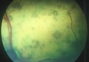
On routine eye examination of this polydypsic polyuric thirteen year old cat you notice these circular lesions in the retinas of both eyes. What is the likely diagnosis, how would you confirm and treat it?
These are small retinal detachments associated with hypertensiono. We call them hypertensive retinopathy but in fact they are a consequence of hypertensive choroidopathy – choroidal vessels are losing fluid which is causing these retinal lesions not associated with the retinal vessels. Here, on the other hand,m is a more advanmced case with retinal haemorrhage and lesions affecting the retinal vessels themselves. Without treatment this case would soon progress to more profound retinal detachment and blindness. Diagnosis would be confirmed by measuring the blood pressure, and treatment should be byt use of oral amlodipine at a dose of 0.625mg daily. That strange dose rate comes because its an eighth of a 5mg tablet which resolves most cat’s hypertension well. Assessment and control of the underlying cause, be it renal failure or hyperthyroidism, is of course important. Read Crispin and Mould’s excellent review for more details: Systemic hypertensive disease and the feline fundus.
Vet Ophthalmol 2001 4:131-40.

You must be logged in to post a comment.