A Clinical Case from the Archives : 17/10/2004

[tab name=”The Case”]This cat is presented emaciated at a charity clinic. It has been rescued from a ramshackle farm where the numerous dogs and cats there are fed the same cheap commercial dog food. What is this retinal lesion and can you explain why the cat is suffering from it?[/tab][tab name=”David’s view”]This is a focal area of retinal degeneration dorolateral to the optic disc. This is the area centralis, a region of highly concentrated photoreceptors with consequently a high energy demand. The retinal degeneration is most likely here to be caused by taurine deficiency. Cats require this amino acid while dogs do not, thus a cat fed on a dog food diet will develop lesions such as this retinopathy and cardiomyopathy. The condition was first identified more than a quarter of a century ago (Hayes et al Retinal degeneration associated with taurine deficiency in the cat.
Science. 1975 188:949-51).
[/tab][end_tabset] 

Posted in Cases | Tagged , | Leave a comment

A Clinical Case from the Archives : 17/10/2004

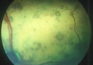
[tab name=”The Case”]On routine eye examination of this polydypsic polyuric thirteen year old cat you notice these circular lesions in the retinas of both eyes. What is the likely diagnosis, how would you confirm and treat it?[/tab][tab name=”David’s view”]These are small retinal detachments associated with hypertensiono. We call them hypertensive retinopathy but in fact they are a consequence of hypertensive choroidopathy – choroidal vessels are losing fluid which is causing these retinal lesions not associated with the retinal vessels. Here, on the other hand,m is a more advanmced case with retinal haemorrhage and lesions affecting the retinal vessels themselves. Without treatment this case would soon progress to more profound retinal detachment and blindness. Diagnosis would be confirmed by measuring the blood pressure, and treatment should be byt use of oral amlodipine at a dose of 0.625mg daily. That strange dose rate comes because its an eighth of a 5mg tablet which resolves most cat’s hypertension well. Assessment and control of the underlying cause, be it renal failure or hyperthyroidism, is of course important. Read Crispin and Mould’s excellent review for more details: Systemic hypertensive disease and the feline fundus.
Vet Ophthalmol 2001 4:131-40.[/tab][end_tabset] 

Posted in Cases | Tagged , , | Leave a comment

A Clinical Case from the Archives : 10/10/2004

[tab name=”The Case”]This three year old cockateil is presented with these ocular signs. What are you seeing, what might be the cause and how would you treat the bird?[/tab][tab name=”David’s view”]The chemosis and conjunctivitis with periorbital swelling and epiphora are characteristic signs of a condition in caged birds which has been seen for many years but in which no infective organism has generally been isolated by conventional methods of bacteriological culture. The condition responds to tetracycline therapy and more recently has been shown in many cases to be caused by Mycoplasma gallisepticum. In the United States a pandemic of the condition is occurring as investigated by several groups of veterinary ophthalmologists (Kollias et al Experimental infection of house finches with Mycoplasma gallisepticum.
J Wildl Dis. 2004 40(1):79-86, Farmer et al, Mycoplasmal conjunctivitis in songbirds from New York, J Wildl Dis. 2000 36(2):257-64. Donht et al, Epidemic mycoplasmal conjunctivitis in house finches from eastern North America.
J Wildl Dis. 1998 34(2):265-80, Fisher et al, Mycoplasmal conjunctivitis in wild songbirds: the spread of a new contagious disease in a mobile host population.
Emerg Infect Dis. 1997 3(1):69-72)). In many of these birds systemic infection is fatal but in caged birds tetracycline therapy is curative.
[/tab][end_tabset] 

Posted in Cases | Tagged , | Leave a comment

A Clinical Case from the Archives : 10/10/2004

[tab name=”The Case”]This three week old snowy owl chick is presented to you blind, these lesions having been present throughout its short life. What might your differential diagnosis list be?[/tab][tab name=”David’s view”]My first thought was that this was a very pronounced vascular keratitis possibly caused by an infection within the egg. But on closer examination the vessels moved independently of the underlying globe. In fact the nictitating membrane was tightly adhered to the conjunctiva on the opposite side of the globe, in a form of symblepharon. This was associated with a septicaemia – read more about the case in our paper Symblepharon with aberrant protrusion of the nictitating membrane in the snowy owl (Nyctea scandiaca). Veterinary Ophthalmology 6(1):11-3, 2003 which you can download on the recent publications page of this website.[/tab][end_tabset] 

Posted in Cases | Tagged , | Leave a comment

A Clinical Case from the Archives : 10/10/2004

[tab name=”The Case”]What is causing the lesions around this budgerigar’s eyes and how would you treat them?[/tab][tab name=”David’s view”]These are classic excresences caused by Knemidocoptes pillae, a mite which causes hyperketatotic growths termed ‘scaly face’ and ‘tassle foot’ by aviculturalists. The picture here, taken many years ago by the great avian veternary surgeon Leslie Arnall, shows a very pronounced case. Treatment is by topical application of ivermectin over several weeks.[/tab][end_tabset] 

Posted in Cases | Tagged , | Leave a comment

A Clinical Case from the Archives : 29/09/2004

[tab name=”The Case”]On examination of this eye you simply cannot find a pupil through which to examine the retina. What is going on, what might have caused it and what treatment might be worthwhile?[/tab][tab name=”David’s view”]Ophthalmologists woud say that in this eye the pupil has been lost. It suggests severe inflammation in which the edges of a tiny miotic pupil have simply fused together. Notice the dark iris and the white masses – probably old fibrin clots. In fact the globe is considerably smaller than the contralateral eye and the intraocular pressure is only 3mmHg while the pressure in the other eye is a normal 15mmHg. The eye is undergoing phthysis and no return to vision is possible. Anti-inflammatory medication to ensure freedom from pain is important as are antibiotics to maintain orbital sterility.[/tab][end_tabset] 

Posted in Cases | Tagged , | Leave a comment

A Clinical Case from the Archives : 29/09/2004

[tab name=”The Case”]What are the abnormalities in this eye? Might they be connected?[/tab][tab name=”David’s view”]Working logically, the first thing to note is the upper eyelid defect with loss of the normal eyelid margin and resultant trichiasis with hair abrading the cornea – possibly post-traumatic but more likely a congenital defect. The other obvious lesion is the corneal ulcer – here made more obvious after the administration of topical fluorescein. It may be that the eyelid margin defect has led to the ulcer, or at the very least prevented it from healing even it is hasn’t caused it in the first place. Topical antibiotis and ocular surface protection with a contact lens might well be appropriate therapeutic measures. Note also that the iris is dark and there is a synechium and a hazy pupil with aqueous flare (difficult to see on this picture I’ll admit). Those signs suggest uveitis probably consequent to corneal irritation. Treatment with topical NSAID (not steroid as there is an ulcer) and atropine would also be worthwhile.[/tab][end_tabset] 

Posted in Cases | Tagged , | Leave a comment

A Clinical Case from the Archives : 29/09/2004

[tab name=”The Case”]This 10 year old cob was presented having failed a vetting because of this strange lesion, symmetrical in both eyes. The referring veterinary surgeons suspected previous uveitis. Would you pass the horse?[/tab][tab name=”David’s view”]The simple answer is ‘yes’! The horse has a very unusual congenital malformation in development of the iris, leaving it with two pupils. The key difference from a uveitis case, as in this horse rightly failed at a vetting, is that the iris in the first horse is light brown and there are no signs of previous inflammation – the band of tissue is not a synechium which would appear much more irregular.[/tab][end_tabset] 

Posted in Cases | Tagged , | Leave a comment

A Clinical Case from the Archives : 28/09/2004

[tab name=”The Case”]This eight year old cat is presented to you as the observant owner had noticed ‘something different’ in the appearance of the left eye. What are the changes that have changed the appearance of this eye, what is the condition and what diagnotic and therapeutic steps might you take? Incidentally the owner is a young lady with two young children and another on the way – does that influence what you might test for and what you might advise?[/tab][tab name=”David’s view”]The abnormalities include raised areas in the iris, which are lymphoid follicles, engorged and proliferating blood vessels in the iris, so-called rubeosis iridis, and grey circular lesions on the interior face of the cornea. These, seen in more detail here, are keratic precipitates (KPs), clusters of lymphoid cells deposited on the inferior corneal endothelium. In fact these lesions are so-called mutton fat KPs which may well be macrophages not lymphoid cells. There are some finer KPs higher up the cornea which are more likely to be lymphocytic. The retina cannot be visualised in this eye, as there is a vitreous flare of inflammatory cells, while the right eye is unaffected. Tonometry of both eyes showed the right to have an intraocular pressure of 18mmHg while the affected left eye had a pressure of only 13mmHg, confirming the diagnosis of uveitis. Numerous infectious agents may be involved from FHV-1, FIP and Bartonella henslae to FeLV, FIV and Toxoplasma. As these last three are most likely to be involved in this case serology for these would be valuable – assessing the Goldmann-Witmer coefficient (the ratio of aqueous to serum antibody titres) can be helpful but needs relatively invasive aqueous paracentesis. The big worry there is Toxoplasma gondii which, being a zoonosis, could be passed on to the children or the baby in utero – stringent hygiene precautions are essential.[/tab][end_tabset] 

Posted in Cases | Tagged , | Leave a comment

A Clinical Case from the Archives : 28/09/2004

[tab name=”The Case”]This eight year old German Shepherd Dog has been acquired from a rescue centre but is brought to you because of this depigmentation of the nictitating membrane. What is the condition and is it a problem for the animal? What treatment might you recommend?[/tab][tab name=”David’s view”]This is classic lymphocytic plasmacytic conjuctivitis commonly seen in this breed of dog. It causes no irritation and no visual disturbance unless the dog has concurrent chronic superficial keratitis. Nevertheless its appearance concerns owners and thus treatment with topical steroid (dexamethasone alcohol in Maxidex or prednisolone acetate in Pred Forte) or topical cyclosporine (Optimmune) can be useful. The nodules seen in the conjunctiva are lymphoid follicles, as demonstrated in this histopathological section.[/tab][end_tabset] 

Posted in Cases | Tagged , | Leave a comment